医用画像工学_backup
2023.6.29.(木) 12:14
医用画像工学
Medical Imaging Engineering
医用画像工学
Medical Imaging Engineering
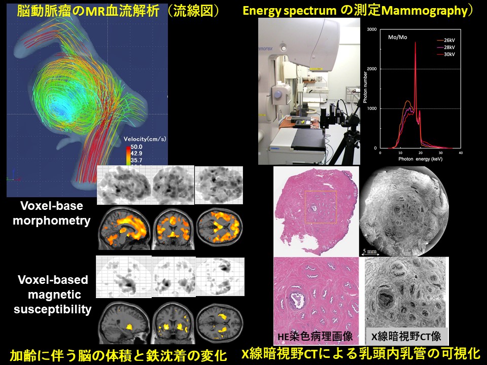
礒田研究室では、磁気共鳴(magneticresonance,MR)による脳機能解析、MRを用いた脳動脈瘤血流解析、MR撮像技術・MR画像診断などの研究を中心に行っています。
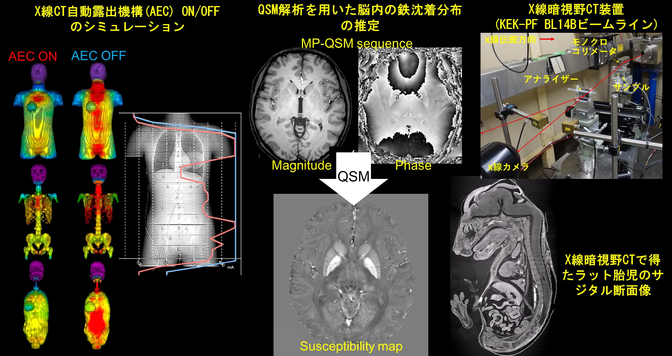
小山研究室は診断領域X線検査による患者被ばく線量の計測方法や評価方法について研究しています.研究では,実測と合わせて,汎用モンテカルロコードElectronGammaShower5(EGS5)による計算を行い,両者による詳細な情報を引き出しています.さらに,外部の企業との共同研究で,エネルギースペクトル情報を利用したエネルギー弁別型フォトンカウンティングマンモグラフィ装置の開発も行っています.
砂口研究室では、生体組織内を高コントラスト・10-6mの分解能で可視化できる、位相コントラストに基づくX線CTの開発を行っており、基礎医学の発展に寄与する新しい医用画像の取得を目的としています。具体的には、高画質化・撮像時間短縮・被曝量削減のための新しい撮像装置や画像処理法の開発に取り組んでいます。近年では、「高エネルギー加速器研究機構」や「知の拠点あいち」といった特別なX線を使用できる放射光施設に撮像システムを整え、乳頭温存乳腺全摘術のリスク低減につながる乳頭内乳管癌の発生メカニズムの解明など、生体のマイクロアナトミーに関する医学的な研究も進めています。
菅研究室では、脳の体積,ミエリン画像,定量的磁化率画像,拡散スペクトル解析についてMRを中心とした画像処理,解析法の研究を行っています.
The Isoda Laboratory mainly focuses on research on brain functional magnetic resonance imaging (fMRI), magnetic resonance fluid dynamics (MRFD) of intracranial aneurysms, magnetic resonance (MR) imaging technology and MR diagnostic imaging.
The Koyama Laboratory is investigating measurement and estimation methods of patient exposure in diagnostic X-ray examinations. In the research, along with actual measurements, calculations are performed using Monte Carlo universal codes, and detailed information is derived from both. Furthermore, we are also developing an energy-resolved photon-counting mammography unit using energy spectrum information in collaboration with a company.
The Sunaguchi Laboratory has been developing a phase-contrast based X-ray CT system that can visualize the inside of biological tissues with high contrast and a spatial resolution of 1 μm, with the aim of acquiring new medical images that will contribute to the development of basic medicine. Especially, we are working on the development of new imaging devices and image processing methods to improve image quality, shorten imaging time, and reduce radiation exposure. In recent years, we have been conducting medical research on microanatomy, such as the elucidation of the developmental mechanism of ductal carcinoma in the nipple, which may reduce the risk of nipple sparing mastectomy, by installing imaging systems in a synchrotron radiation facility such as KEK-PF or AichiSR.
The Kan Laboratory conducts studies of medical image processing and analysis on magnetic resonance imaging, regarding brain volume, myelin water fraction map (MWF), quantitative susceptibility map (QSM), and diffusion spectrum analysis in the brain.
基礎放射線技術学
2020.6.4.(木) 14:27

基礎放射線技術学
Basic Radiological Technology
基礎放射線技術学
Basic Radiological Technology


医用画像解析学
2020.6.4.(木) 14:26
医用画像解析学
Medical Imaging Analysis
医用画像解析学
Medical Imaging Analysis
Our team is investigating the medical image processing and analysis and the evaluation of radiation doses in order to make them compatible with each other, by incorporating modern mathematical technology. Each of our team is working hard on various unique and ingenious research projects and aiming at getting good research results.
生体機能科学
2020.6.4.(木) 14:26

生体機能科学
Biofunctional Sciences
生体機能科学
Biofunctional Sciences
編集中
医用画像工学
2020.6.4.(木) 14:25
医用画像工学
Medical Imaging Engineering
医用画像工学
Medical Imaging Engineering
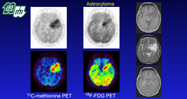
西井研究室では「基礎から臨床応用にいたる画像研究を行い、新しい医用画像を患者にとどける!」モットーに活動しています。I)分子の動きとらえ、II)体のしくみや病態を画像化し、III)臨床現場で実践する研究、さらにがん治療に直結するTheranostics 研究(Therapy + Diagnosis)も展開しています。
1. アミノ酸やチミジン誘導体などを用いた新しいPET画像診断のトランスレーショナル研究と臨床応用研究。
2. 治療前後の定量的画像解析によるがん個別化医療に向けた臨床画像診断法の開発。
3. 標的アイソトープ治療による核医学がん治療の開発と、その安全で効果的な実践に向けた線量シミュレーション研究。
4. AIやICT技術を応用した新しい画像診断研究。
健康長寿社会の実現を達成するために、放射線画像や治療技術を活用した医療イノベーションの創出と、医療ニーズやグローバル化に対応できる研究を目指しています。
小山研究室は診断領域X線検査による患者被ばく線量の計測方法や評価方法について研究しています.研究では,実測と合わせて,汎用モンテカルロコードElectron Gamma Shower5(EGS5)による計算を行い,両者による詳細な情報を引き出しています.さらに,外部の企業との共同研究で,エネルギースペクトル情報を利用したエネルギー弁別型フォトンカウンティングマンモグラフィ装置の開発も行っています.
砂口研究室では、生体組織内を高コントラスト・数マイクロメートルの分解能で可視化できる、位相コントラストに基づくX線CTの開発を行っており、基礎医学の発展に寄与する新しい医用画像の取得を目的としています。具体的には、高画質化・撮像時間短縮・被曝量削減のための新しい撮像装置や画像処理法の開発に取り組んでいます。近年では、「高エネルギー加速器研究機構」や「知の拠点あいち」といった特別なX線を使用できる放射光施設に撮像システムを整え、乳頭温存乳腺全摘術のリスク低減につながる乳頭内乳管癌の発生メカニズムの解明など、生体のマイクロアナトミーに関する医学的な研究も進めています。
菅研究室では、脳の体積,ミエリン画像,定量的磁化率画像,拡散スペクトル解析についてMRを中心とした画像処理,解析法の研究を行っています.
The mission of Nishii Laboratory is “To bring new medical images to patients through imaging research from basic to clinical applications!” Our research focuses on (i) understanding molecular behavior, (ii) visualizing the mechanisms and pathophysiology inside the body, and (iii) applying them to clinical practice, through basic science, preclinical models and clinical trials. In addition, our research spans theranostics research (Therapy + Diagnosis), which is directly associated with cancer treatment.
Our current research projects focus on:
1. translational research and clinical trials of novel PET imaging.
2. quantitative clinical imaging for individualized cancer treatment.
3. targeted radionuclide therapy (TRT).
4. AI-based imaging
The Koyama Laboratory is investigating measurement and estimation methods of patient exposure in diagnostic X-ray examinations. In the research, along with actual measurements, calculations are performed using Monte Carlo universal codes, and detailed information is derived from both. Furthermore, we are also developing an energy-resolved photon-counting mammography unit using energy spectrum information in collaboration with a company.
The Sunaguchi Laboratory has been developing a phase-contrast based X-ray CT system that can visualize the inside of biological tissues with high contrast and a spatial resolution of 1 μm, with the aim of acquiring new medical images that will contribute to the development of basic medicine. Especially, we are working on the development of new imaging devices and image processing methods to improve image quality, shorten imaging time, and reduce radiation exposure. In recent years, we have been conducting medical research on microanatomy, such as the elucidation of the developmental mechanism of ductal carcinoma in the nipple, which may reduce the risk of nipple sparing mastectomy, by installing imaging systems in a synchrotron radiation facility such as KEK-PF or AichiSR.
The Kan Laboratory conducts studies of medical image processing and analysis on magnetic resonance imaging, regarding brain volume, myelin water fraction map (MWF), quantitative susceptibility map (QSM), and diffusion spectrum analysis in the brain.
医用量子科学
2020.6.4.(木) 14:24
医用量子科学
Medical Quantum Science
医用量子科学
Medical Quantum Science
In this “Medical Quantum Science” course, the members are conducting our researches mainly on radiotherapies. In radiation therapies, high energy X-ray and electrons, particle therapies, and brachytherapy are now roughly used and we are developing new technologies for these fields. Moreover we are also intensively exploring the emerging fields of medical quantum science to be the top of the world in this research field.
Electron Gamma Shower Version 5 (EGS5)の利用と電子線付加フィルタの開発(小口研究室)
放射線の物質内の相互作用をシミュレーションする汎用モンテカルロ計算コードのひとつにElectron Gamma Shower Version 5 (EGS5)があります。また、複雑な計算ジオメトリを構築するためにGUIソフトウェアの1つであるCGViewを用いることで、より複雑なモデルでのシミュレーションが可能となります。CGViewによって治療用直線加速器のヘッド構造を忠実に再現し、EGS5により加速器ターゲットより射出された粒子 (光子・電子・陽電子)のヒストリーを詳細に計算し、使用されている物質情報の記述を行うことで自由度の高い計算を精度良く行うことが可能となります。単純な条件でシミュレーション結果を実測値と比較しその整合性を検証することにより、実測が難しい領域でのシミュレーション計算精度が担保できます。
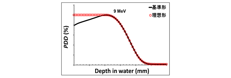
当研究室では高エネルギー電子線治療における深部線量分布 (PDD)の最適化を図る付加フィルタの開発を行っています。電子線はX 線やγ線と異なり、荷電粒子であるため物質中を通過する際に周辺物質にエネルギーを落とす相互作用をしつづけ、その運動エネルギーがなくなると停止して消滅します。その結果、高エネルギー電子線による物質内の線量分布はある深さで線量が最大となり、その後深部で急激に減少する特徴を示します(図1)。この特徴により、電子線は主に表在性腫瘍の治療に用いられていますが、エネルギーが低下するにつれて入射表面(浅部)の線量もより低下してしまう特性も持ち合わせています。
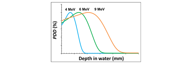
図1 高エネルギー電子線(4, 6, 9 MeV)の水中における深部線量分布
理想的には浅部領域の腫瘍に対して腫瘍全範囲で高線量を保ち、腫瘍後方の正常組織では線量が激減する分布が望まれます(図2)。そこで、本研究ではエネルギーが低下した場合でも飛程を保ちつつ、浅部線量を高く補償するような付加フィルタの開発をおこなっています。私たちはこのフィルタの設計にEGS5を利用しています。
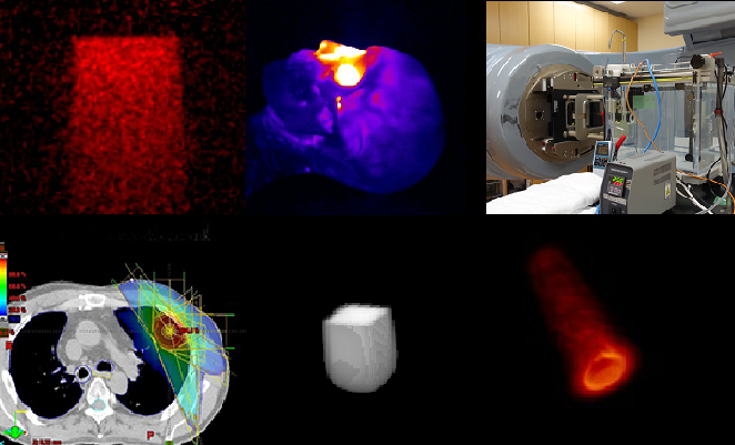
ポリマーゲル線量計の開発(小口研究室)
3次元線量分布を測定するための線量計として、現在、ポリマーゲル線量計の研究を行っています。ポリマーゲル線量計は化学線量計の1つで、ビニルモノマーの放射線重合反応を利用しています。我々が作製しているのはMethacrylic and Ascorbic acid in Gelatin Initiated by Copper (MAGIC)ゲル線量計という種類のもので、その組成を表1に示します。

ポリマーゲル線量計に放射線が照射されると、放射線重合反応によりモノマーがポリマーとなり析出します(図1)。モノマーが重合反応しポリマーとなる量は吸収線量に比例します。したがって、ポリマーの量を測定することで吸収線量の計測ができます。モノマーがポリマーになると、周囲の水分子の環境や物理的密度、光の透過率が変化します。そのため、ポリマー量はmagnetic resonance (MR)やX線 computed tomography (CT)、optical CTで測定できます。ポリマーゲル線量計での計測は、信号値と吸収線量を結びつける校正曲線作成過程と線量分布を取得するファントム過程に分けられます。ファントム検証で取得された信号強度分布を作成した校正曲線で線量分布に変換することで、ファントム内の線量分布測定が可能となります。
容器の形を変えれば、患者を模擬したポリマーゲル線量計を作製することができます。これにより、実際の放射線治療時に近い患者体内線量分布が取得可能であると考えられます。しかし、ポリマーゲル線量計の特性は、組成、作製手順、ファントムの大きさ、保管温度、読み取り時のパラメータなどの影響を受けます。そこで、我々は、組成や保管、撮像の条件などを変えて、より良いポリマーゲル線量計の作製および読取りを目指し研究しています。

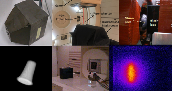
炭素線治療における中性子被ばくの低減化に関する研究(小森研究室)
高エネルギーに加速された陽子または炭素イオンを用いた粒子線がん治療は、標的腫瘍への線量集中性がよく、障害が少ない治療が可能です。粒子線治療における照射野形成法の一つである拡大照射法は、シンクロトロンなどの加速器から供給された直径1cm程度のビームを、横・深さ方向に拡大して線量が一様な照射野を形成し標的腫瘍に照射します。拡大照射法のデメリットの一つとして、中性子被ばくがあげられます。粒子線が照射野形成装置と衝突すると二次中性子が発生します。照射野形成装置は患者さんの直前に設置する必要がありますので、不要な被ばくを受けることになります。もちろん、がんを治療するメリットの方がはるかに大きいので、拡大照射法を用いて粒子線を患者に照射しますが、不要な被ばくは少ないにこしたことはありません。
我々はモンテカルロシミュレーションを用いて、照射野形成装置に中性子被ばく低減に最適化されたコリメータを追加配置することで、どの程度の低減効果があるのかを検証しました。

放射線医学総合研究所の炭素線治療装置HIMACの照射野形成装置をシミュレーション内に構築し、コリメータの位置、大きさ、材質などの違いによる、患者位置での中性子周辺線量当量の変化を計算しました。
結果の一例を図1に示します。横軸は標的腫瘍からの距離、縦軸は照射線量当たりの中性子周辺線量当量を示します。最適な場所にコリメータを追加することで、35%程度の低減が確認されました。
研究成果は下記学会で発表を行いました。現在、海外雑誌に論文を投稿中です。
Monte Carlo study on reduction in the secondary neutron exposure in passive carbon-ion radiotherapy. European Congress of Radiology 2013; EPOS™ Scientific Exhibition. 2013
モンテカルロシミュレーションを用いたパッシブ炭素線治療時の二次中性子被ばくの低減. 第105回日本医学物理学会学術大会. 2013
モンテカルロシミュレーションによる炭素線治療における二次中性子線量の低減に関する検討. 第5回中部放射線医療技術学術大会. 2012
医用機能画像評価学
2020.6.4.(木) 14:24
医用機能画像評価学
Functional Medical Imaging
医用機能画像評価学
Functional Medical Imaging
核医学は、放射性同位元素(radioisotope:RI)を利用して診断や治療を行う医学です。放射線医学を構成する主要な分野であり,臓器の機能・代謝を知る方法として重要です。とりわけ単一光子横断断層像(SPECT、single photon emission computed tomography),ポジトロン横断断層像(PET、positron emission tomography)を用いる施設も増加し,この分野における進歩はめざましいものがあります。また、核医学において、生体内でおこる様々な事象を標的とする放射性医薬品の開発や、その性質を明らかにすることは診断・治療の両面において重要な役割を果たしています。医用機能画像評価学ユニットでは、核医学の基礎から臨床まで幅広い領域の研究に取り組むとともに、大学院生、学部学生の教育にも力を注いでいます。
In nuclear medicine, treatment and diagnosis of diseases are performed by means of administering radioisotopes (RI). Nuclear medicine is one of the major fields of radiology and it provides important tools to analyze the functions and metabolism of organs. In particular, the facilities equipped with single-photon emission tomography (SPECT) and positron emission tomography (PET) are increasing, and there is a remarkable improvement in imaging technologies using SPECT and PET. The development and characterization of radiopharmaceuticals or molecular probes are also playing critical roles in nuclear medicine. In Functional Medical Imaging Unit, we pursue clinical studies along with the basic studies with imaging techniques of nuclear medicine and radiopharmaceuticals, and we put major emphasis on education of postgraduate and undergraduate students.
基礎放射線技術学
2016.3.22.(火) 02:58
基礎放射線技術学
Basic Radiological Technology
基礎放射線技術学
Basic Radiological Technology
生体機能科学
2016.3.22.(火) 02:57
生体機能科学
Biofunctional Sciences
生体機能科学
Biofunctional Sciences
編集中
医用画像工学
2016.3.22.(火) 02:55
医用画像工学
Medical Imaging Engineering
医用画像工学
Medical Imaging Engineering
小山研究室は診断領域X線検査による患者被ばく線量の計測方法や評価方法について研究しています.研究では,実測と合わせて,汎用モンテカルロコードElectronGammaShower5(EGS5)による計算を行い,両者による詳細な情報を引き出しています.さらに,外部の企業との共同研究で,エネルギースペクトル情報を利用したエネルギー弁別型フォトンカウンティングマンモグラフィ装置の開発も行っています.
砂口研究室では、生体組織内を高コントラスト・10-6mの分解能で可視化できる、位相コントラストに基づくX線CTの開発を行っており、基礎医学の発展に寄与する新しい医用画像の取得を目的としています。具体的には、高画質化・撮像時間短縮・被曝量削減のための新しい撮像装置や画像処理法の開発に取り組んでいます。近年では、「高エネルギー加速器研究機構」や「知の拠点あいち」といった特別なX線を使用できる放射光施設に撮像システムを整え、乳頭温存乳腺全摘術のリスク低減につながる乳頭内乳管癌の発生メカニズムの解明など、生体のマイクロアナトミーに関する医学的な研究も進めています。
菅研究室では、脳の体積,ミエリン画像,定量的磁化率画像,拡散スペクトル解析についてMRを中心とした画像処理,解析法の研究を行っています.
The Koyama Laboratory is investigating measurement and estimation methods of patient exposure in diagnostic X-ray examinations. In the research, along with actual measurements, calculations are performed using Monte Carlo universal codes, and detailed information is derived from both. Furthermore, we are also developing an energy-resolved photon-counting mammography unit using energy spectrum information in collaboration with a company.
The Sunaguchi Laboratory has been developing a phase-contrast based X-ray CT system that can visualize the inside of biological tissues with high contrast and a spatial resolution of 1 μm, with the aim of acquiring new medical images that will contribute to the development of basic medicine. Especially, we are working on the development of new imaging devices and image processing methods to improve image quality, shorten imaging time, and reduce radiation exposure. In recent years, we have been conducting medical research on microanatomy, such as the elucidation of the developmental mechanism of ductal carcinoma in the nipple, which may reduce the risk of nipple sparing mastectomy, by installing imaging systems in a synchrotron radiation facility such as KEK-PF or AichiSR.
The Kan Laboratory conducts studies of medical image processing and analysis on magnetic resonance imaging, regarding brain volume, myelin water fraction map (MWF), quantitative susceptibility map (QSM), and diffusion spectrum analysis in the brain.