医用画像工学
Medical Imaging Engineering
西井研究室では「基礎から臨床応用にいたる画像研究を行い、新しい医用画像を患者にとどける!」モットーに活動しています。I)分子の動きとらえ、II)体のしくみや病態を画像化し、III)臨床現場で実践する研究、さらにがん治療に直結するTheranostics 研究(Therapy + Diagnosis)も展開しています。
1. アミノ酸やチミジン誘導体などを用いた新しいPET画像診断のトランスレーショナル研究と臨床応用研究。
2. 治療前後の定量的画像解析によるがん個別化医療に向けた臨床画像診断法の開発。
3. 標的アイソトープ治療による核医学がん治療の開発と、その安全で効果的な実践に向けた線量シミュレーション研究。
4. AIやICT技術を応用した新しい画像診断研究。
健康長寿社会の実現を達成するために、放射線画像や治療技術を活用した医療イノベーションの創出と、医療ニーズやグローバル化に対応できる研究を目指しています。
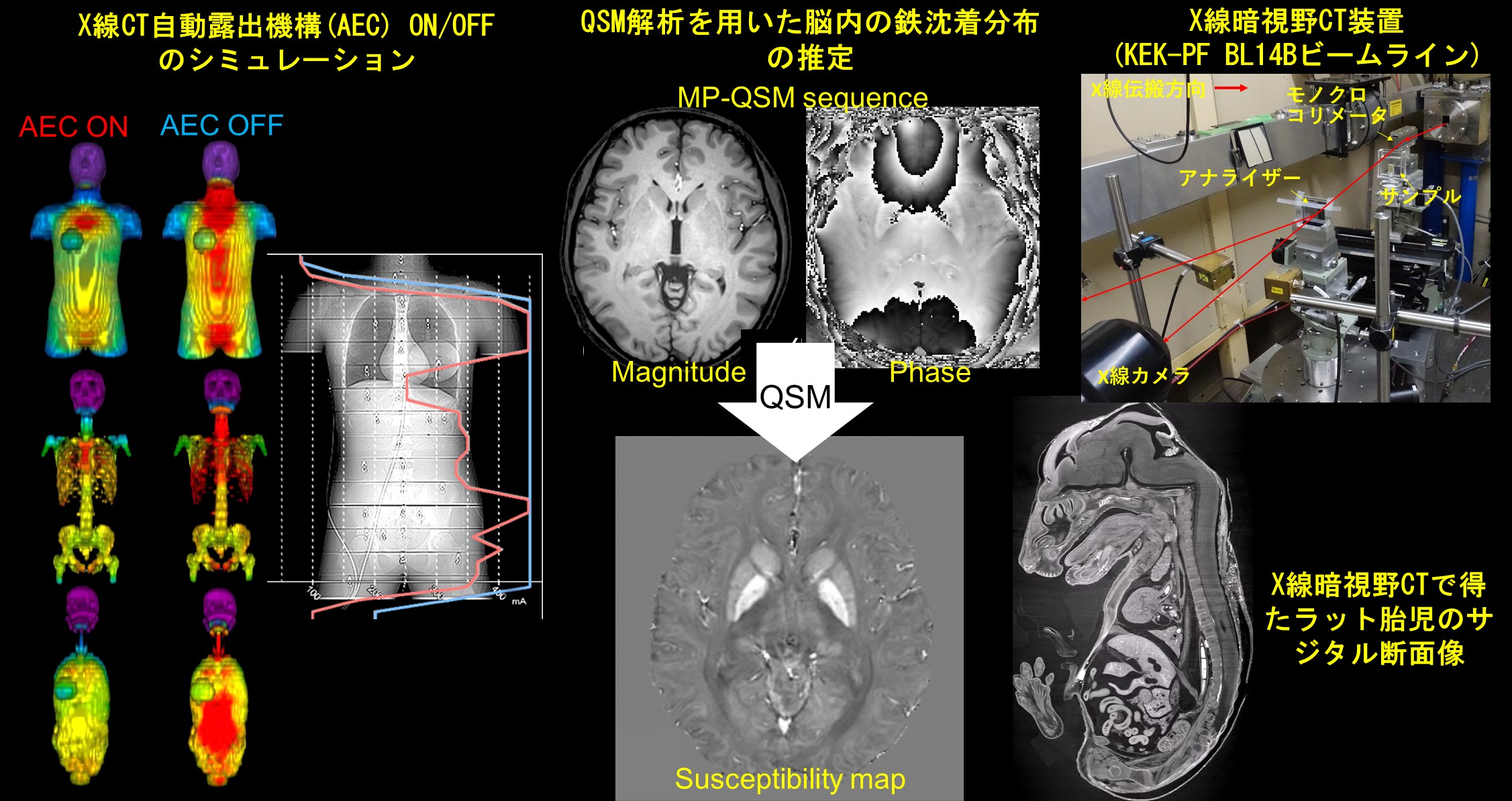
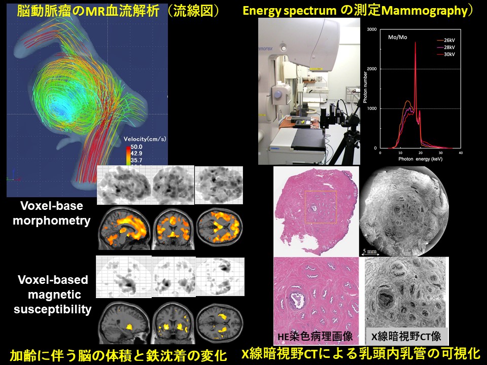
小山研究室は診断領域X線検査による患者被ばく線量の計測方法や評価方法について研究しています.研究では,実測と合わせて,汎用モンテカルロコードElectron Gamma Shower5(EGS5)による計算を行い,両者による詳細な情報を引き出しています.さらに,外部の企業との共同研究で,エネルギースペクトル情報を利用したエネルギー弁別型フォトンカウンティングマンモグラフィ装置の開発も行っています.
砂口研究室では、生体組織内を高コントラスト・数マイクロメートルの分解能で可視化できる、位相コントラストに基づくX線CTの開発を行っており、基礎医学の発展に寄与する新しい医用画像の取得を目的としています。具体的には、高画質化・撮像時間短縮・被曝量削減のための新しい撮像装置や画像処理法の開発に取り組んでいます。近年では、「高エネルギー加速器研究機構」や「知の拠点あいち」といった特別なX線を使用できる放射光施設に撮像システムを整え、乳頭温存乳腺全摘術のリスク低減につながる乳頭内乳管癌の発生メカニズムの解明など、生体のマイクロアナトミーに関する医学的な研究も進めています。
菅研究室では、脳の体積,ミエリン画像,定量的磁化率画像,拡散スペクトル解析についてMRを中心とした画像処理,解析法の研究を行っています.
The mission of Nishii Laboratory is “To bring new medical images to patients through imaging research from basic to clinical applications!” Our research focuses on (i) understanding molecular behavior, (ii) visualizing the mechanisms and pathophysiology inside the body, and (iii) applying them to clinical practice, through basic science, preclinical models and clinical trials. In addition, our research spans theranostics research (Therapy + Diagnosis), which is directly associated with cancer treatment.
Our current research projects focus on:
1. translational research and clinical trials of novel PET imaging.
2. quantitative clinical imaging for individualized cancer treatment.
3. targeted radionuclide therapy (TRT).
4. AI-based imaging
The Koyama Laboratory is investigating measurement and estimation methods of patient exposure in diagnostic X-ray examinations. In the research, along with actual measurements, calculations are performed using Monte Carlo universal codes, and detailed information is derived from both. Furthermore, we are also developing an energy-resolved photon-counting mammography unit using energy spectrum information in collaboration with a company.
The Sunaguchi Laboratory has been developing a phase-contrast based X-ray CT system that can visualize the inside of biological tissues with high contrast and a spatial resolution of 1 μm, with the aim of acquiring new medical images that will contribute to the development of basic medicine. Especially, we are working on the development of new imaging devices and image processing methods to improve image quality, shorten imaging time, and reduce radiation exposure. In recent years, we have been conducting medical research on microanatomy, such as the elucidation of the developmental mechanism of ductal carcinoma in the nipple, which may reduce the risk of nipple sparing mastectomy, by installing imaging systems in a synchrotron radiation facility such as KEK-PF or AichiSR.
The Kan Laboratory conducts studies of medical image processing and analysis on magnetic resonance imaging, regarding brain volume, myelin water fraction map (MWF), quantitative susceptibility map (QSM), and diffusion spectrum analysis in the brain.